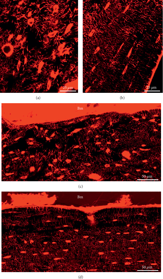
Figure 7.

High-magnification images of OLCN stained with rhodamine and visualized with fluorescence confocal laser scanning microscopy. MM-injected femur with (a) larger, irregular-shaped osteocyte lacunae within a disorganized canaliculi network architecture in proximity to the bone marrow (indicated with # in Figure 3(f)) and (b) flat osteocyte lacunae organized in a lamellar structure on the periosteal side (indicated with ∗ in Figure 3(f)). High-magnification image of the connection of the osteocyte canaliculi network to the bone marrow on the endosteal surface of (c) a MM-injected femur with a disrupted network and (d) a healthy control mouse femur illustrating an organized network in lamellae parallel to the bone surface and canaliculi perpendicular to them.
