
Figure 2:
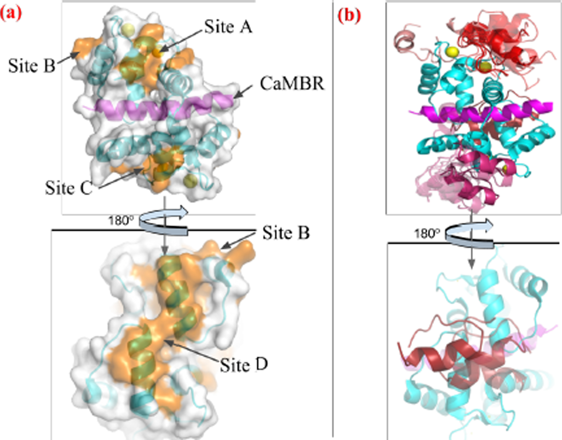
(a) Four tentative binding sites (orange) on the surface of CaM-CaMBR complex. CaM is colored in cyan, CaMBR is colored in magenta and Ca2+ ions are colored in yellow. (b) ZDOCK predicted conformations of the distal helix interacting with CaM/CaMBR complex at each site. Predicted distal helix conformations from site A to D are colored as red, salmon, warmpink and firebrick, respectively.