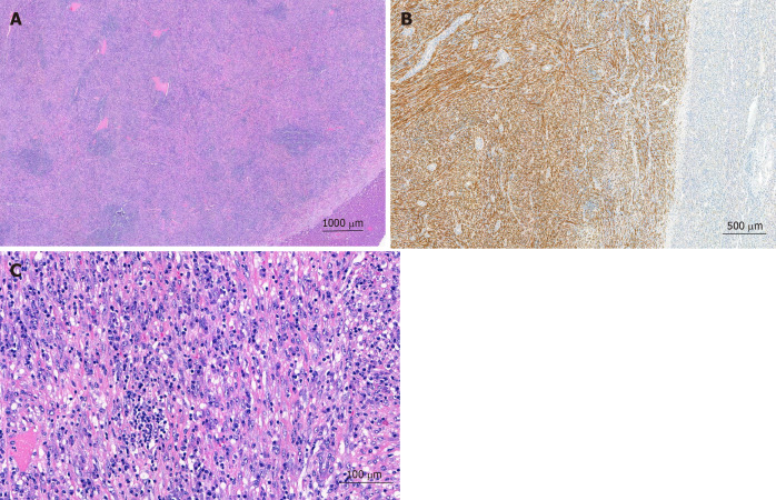
Figure 3.
Postoperative microscopic pathology of the inflammatory myofibroblastic tumors. A: Well demarcated firm vascularized tumor mass with spotty inflammatory infiltrate; B: Bland proliferation of spindle cells in broad fascicles at higher magnification. Scattered lymphocytes and plasma cell; C: Intense positivity of the spindel cells for anaplastic lymphoma kinase.

