Figure 1.
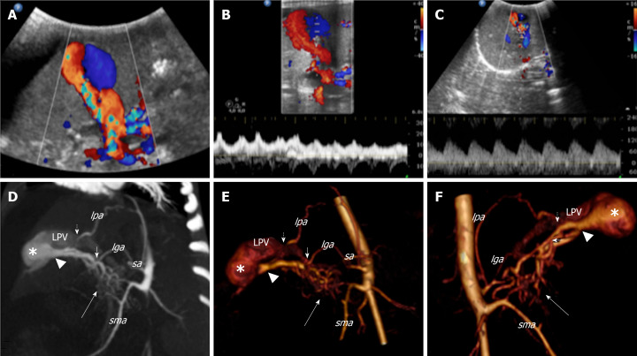
Colour Doppler ultrasound and computed tomography images that show the congenital intrahepatic arterioportal fistula. A: Colour Doppler ultrasound images of the liver revealed aneurysmal dilatation of the left portal vein with turbulent bidirectional flow; B: Particularly, the venous flow appeared arterialised; C: Several dilated and tortuous branches of the hepatic artery with high flow rate supplied the dilated portal vein segment; D: Contrast enhanced multi-detector computer tomography images of the abdomen reformatted on both the left oblique: maximum intensity projection; E: 3D volume rendered; F: 3D-VR right oblique plane. Contrast enhanced multi-detector computer tomography shows the complex vascular malformation characterised by a dense dysplastic small arterial network that arose from the hepatic and superior mesenteric system (thick arrow) and directly converged to the left portal vein through one Y-shaped fistula within the Rex recess (arrow head). Two additional feeders to the vascular anomaly [from the phrenic (short dotted arrow) and left gastric (short arrow) artery, respectively] were also detected. lga: Left gastric artery; lpa: Left phrenic artery; sa: Splenic artery; sma: Superior mesenteric artery; LPV: Left portal vein.

