Figure 3.
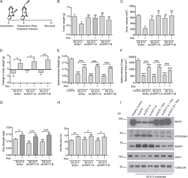
SIRT1 stabilization in muscles regulates muscles wasting in vivo. (A) Schematic illustration of the treatment strategy. (B and C) Postnecropsy measurements of S2-013 shScr (n = 10), shSIRT1-A (n = 10), and shSIRT1-B (n = 10) tumor weights (B) and tumor volumes (C). (D) Change in body weight measurements of the tumor-bearing mice 21 d after implantation. (E) Postnecropsy gastrocnemius muscle weight from S2-013 shScr, S2-013 shSIRT1-A, and S2-013 shSIRT1-B tumor-bearing mice. (F) Quantification of muscle fiber cross-sectional area in hematoxylin and eosin–stained muscle sections from tumor-bearing mice. (G) Measurement of grip strength of the tumor-bearing mice 18 d after implantation. (H) Measurement of fat percentage of S2-013 shScr (n = 8), S2-013 shSIRT1-A (n = 8), and S2-013 shSIRT1-B (n = 8) tumor-bearing mice by dual-energy x-ray absorptiometry scanning 18 d after implantation. (I) Immunoblots of muscle tissue extracts from the tumor-bearing mice treated with resveratrol or solvent control, showing regulation of MyHC, Atrogin-1, MuRF1, and SIRT1. Tubulin was used as a loading control. All in vitro experiments were performed at least three times in triplicate. Data are mean ± SEM compared with one-way ANOVA with Bonferroni’s multiple comparisons (B–H); *, P < 0.05; **, P < 0.01; ***, P < 0.001.

