Figure 4.
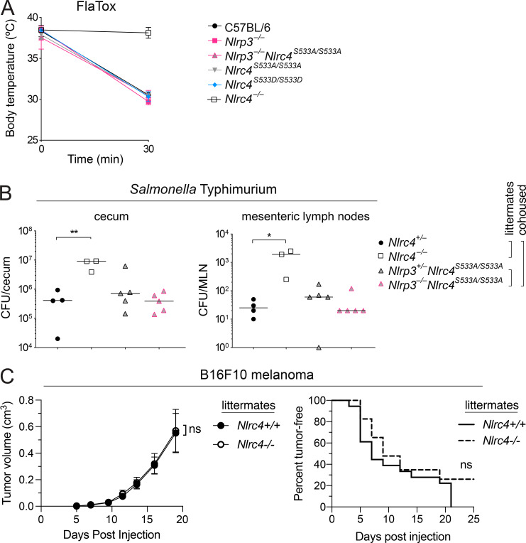
In vivo disease susceptibility of Nlrc4 mutant mice. (A) Rectal temperatures of WT C57BL6/J, Nlrp3−/−, Nlrc4S533A/ S533A;Nlrp3−/−, Nlrc4S533A/S533A, Nlrc4S533D/S533D, and Nlrc4−/− mice (n = 3 per group) injected retroorbitally with 0.8 µg/g body weight of PA and 0.4 µg/g LFn-FlaA. Mean ± SD. (B) CFU in cecum and mesenteric lymph node (MLN) 18 h after oral S. Typhimurium infection of Nlrc4S533A/S533A;Nlrp3−/− and Nlrc4S533A/S533A;Nlrp3−/− littermate mice cohoused with Nlrc4+/− and Nlrc4−/− littermate mice (n = 3–5 per group). Median; Mann–Whitney test, *, P < 0.01, **, P < 0.005. (A and B) Data are representative of three independent experiments. (C) Tumor size and incidence of B16F10 melanoma injected subcutaneously into Nlrc4+/+ and Nlrc4−/− littermate mice (n = 18 or 23, respectively). Mean ± SEM. Data are pooled from three independent experiments. Repeated measures two-way ANOVA with Sidak’s multiple comparisons test (tumor volume) or log-rank Mantel–Cox test (tumor incidence); ns, not significant.

