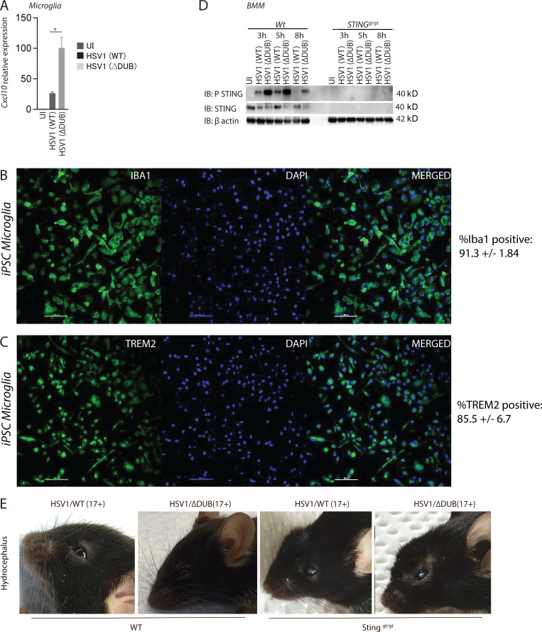
Figure S3.
Activation of IFN responses by DUB-deficient HSV and disease development in vivo. (A) WT primary microglia were isolated and infected with HSV1 (WT) or HSV1 ΔDUB virus (MOI 3) for 16 h. Total RNA was isolated, and Cxcl10 levels were measured and normalized to β-actin by quantitative RT-PCR. (B and C) iPSCs were subjected to differentiation into microglia. The resulting cell population was stained for microglia markers Iba1 and TREM2 and visualized by immunofluorescence. (D) WT and Stinggt/gt bone marrow–derived macrophages (BMMs) were infected with HSV1 (WT) or HSV1 ΔDUB (MOI:10) for the indicated time intervals. Cleared lysates were immunoblotted with the indicated antibodies. (E) C57BL/6 WT and Stinggt/gt mice were infected in the cornea with HSV1 (WT) or HSV1 ΔDUB (17+ strain). The figure shows representative images of mice from each group. P value was calculated using a two-tailed unpaired Student’s t test. *, P < 0.05. Error bars represent standard deviation.

