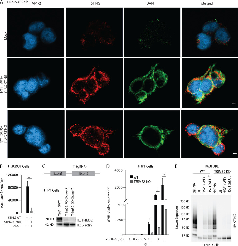
Figure S5.
Localization of VP1-2 NT1 and STING in HEK293T cells and role for STING K150 and TRIM32 for full activation of STING. (A) HEK293T cells were transfected with VP1-2 NT1 and STING. The cells were fixed 24 h after transfection and stained with anti–VP1-2 and anti-STING. Nuclear were visualized by staining with DAPI. Scale bars, 5 µm. (B) HEK293T cells were transfected with (50 ng) FLAG-tagged WT or K150R STING, cGAS, and IFNB1 promoter luciferase reporter and β-actin Renilla reporter. Reporter gene activity was measured 24 h after transfection (n = 3). (C) Illustration of targeting region for gRNA used to generate TRIM32 KO cells. Lysates from WT and two TRIM32 KO clones lysates were immunoblotted with anti-TRIM32 and anti–β-actin. (D) Shorter exposure of the anti-STING immunoblot shown to the right in Fig 6 F. (E) Type I IFN bioactivity levels in supernatants from PMA-differentiated THP1 (WT), and TRIM32 KO cells were transfected with dsDNA (0.25–5 µg) for 8 h. *, P < 0.05; **, P < 0.01; ns, not significant. Error bars represent standard deviation.

