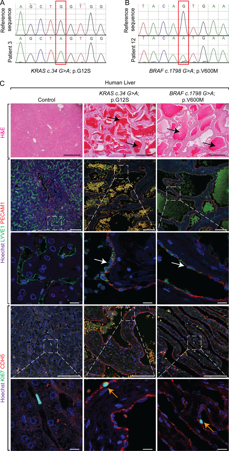
Figure 1.
Human patients with hepatic vascular cavernomas exhibit somatic gain-of-function mutations in KRAS or BRAF genes in pathological liver tissue samples. (A and B) Sanger sequencing of human hepatic vascular cavernoma tissue samples identified a somatic mutation (red box), c.34G>A (p.G12S) in KRAS (A) or c.1798G>A (p.V600M) in BRAF (B) genes. (C) H&E staining on pathological human liver sections shows cavernous spaces filled with erythrocytes (black arrows; n = 14). Coimmunofluorescent staining with Hoechst nuclear counterstain (blue) shows contribution of LYVE1+ (green) and PECAM1+ (red) endothelial cells (white arrows) to hepatic cavernous vascular malformations (n = 7). KI67 (green) and CDH5 (red) coimmunofluorescent staining with Hoechst nuclear counterstain (blue) on pathological human liver sections shows normal proliferation of CDH5+ sinusoidal endothelial cells lining cavernous vascular malformations (orange arrows; control, n = 4; KRASG12/13Mut, n = 11; BRAFV600M, n = 3). All experimental data verified in at least two independent experiments. Scale bars: 500 µm (top row), 100 µm (second and fourth rows from top), and 10 µm (third and fifth rows from top).

