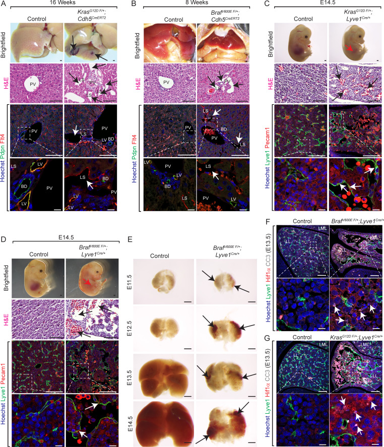
Figure 2.
Endothelial KrasG12D or BrafV600E gain-of-function mutations cause hepatic vascular cavernomas in mice. (A and B) Dissected livers and H&E-stained liver frontal sections show vascular cavernomas (black arrows) in tamoxifen-treated KrasG12D F/+; Cdh5CreERT2 (A) and BrafV600E F/+; Cdh5CreERT2 (B) mice (n = 3). Pdpn (green) and Flt4 (red) coimmunofluorescent staining with Hoechst nuclear counterstain (blue) on KrasG12D F/+; Cdh5CreERT2 (A) and BrafV600E F/+; Cdh5CreERT2 (B) liver sections shows contribution of Flt4+ and Pdpn-negative endothelial cells to hepatic vascular cavernomas (white arrows). n = 3. Scale bars: 500 µm (top row), 100 µm (second and third row from top), and 10 µm (bottom row). Two independent experiments. (C and D) Dissected KrasG12D F/+; Lyve1Cre (C) and BrafV600E F/+; Lyve1Cre (D) embryonic day 14.5 (E14.5) embryos show reduced liver size (red arrow; n = 3). H&E-stained KrasG12D F/+; Lyve1Cre (C) and BrafV600E F/+; Lyve1Cre (D) E14.5 liver frontal sections show vascular cavernomas (black arrows; n = 3). Coimmunofluorescent staining with Hoechst nuclear counterstain (blue) shows contribution of Lyve1+ (green) and Pecam1+ (red) endothelial cells (white arrows) to hepatic vascular cavernomas. n = 3. Scale bars: 500 µm (top row), 100 µm (second and third row from top), and 10 µm (bottom row). Two independent experiments. (E) Dissected BrafV600E F/+; Lyve1Cre livers show reduction in liver size at E13.5 and progressive development of hepatic vascular cavernomas (black arrows). n = 3. Scale bars: 500 µm. (F) Lyve1 (green), Hif1α (red), and Cleaved caspase 3 (CC3, white) coimmunofluorescent staining with Hoechst nuclear counterstain (blue) on BrafV600E F/+; Lyve1Cre (F) and KrasG12D F/+; Lyve1Cre (G) E13.5 liver frontal sections shows hypoxia and apoptosis in clusters of nonendothelial hepatic cells near vascular cavernomas (white arrows). n = 3. Scale bars: 100 µm (top row) and 10 µm (bottom row). Two independent experiments. Littermates were used as controls for all experiments. All experimental data verified in at least two independent experiments. BD, bild duct; LL, left lobe; LML, left medial lobe; LS, liver sinusoid; LV, lymphatic vessel; PV, portal vein.

