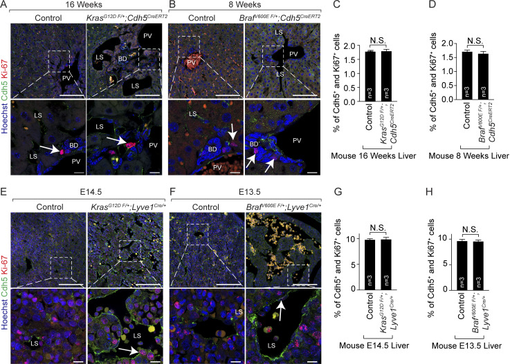
Figure S3.
Mice with endothelial KrasG12D or BrafV600E gain-of-function mutations exhibit normal proliferation of hepatic endothelial cells. (A and B) Ki67 (red) and Cdh5 (green) coimmunofluorescent staining with Hoechst nuclear counterstain (blue) on tamoxifen-treated KrasG12D F/+; Cdh5CreERT2 (A) and BrafV600E F/+; Cdh5CreERT2 (B) adult murine liver sections. White arrows show Ki67+ cells. n = 3. Scale bars: 100 µm (top row) and 10 µm (bottom row). Two independent experiments. FDR, false discovery rate. (C and D) Quantitation of proliferating Cdh5+ cells in tamoxifen-treated control, KrasG12D F/+; Cdh5CreERT2 (C), and BrafV600E F/+; Cdh5CreERT2 (D) embryonic murine livers. n = 3. Unpaired t test, P > 0.8 (C) and P > 0.4 (D). Two independent experiments. Data represent the mean ± SEM. (E and F) Ki67 (red) and Cdh5 (green) coimmunofluorescent staining with Hoechst nuclear counterstain (blue) on KrasG12D F/+; Lyve1Cre/+ (C) and BrafV600E F/+; Lyve1Cre/+ (D) embryonic murine liver sections. White arrows show Ki67+ cells. n = 3. E, embryonic day. Scale bars: 100 µm (top row) and 10 µm (bottom row). Two independent experiments. (G and H) Quantitation of proliferating Cdh5+ cells in control, KrasG12D F/+; Lyve1Cre/+ (G), and BrafV600E F/+; Lyve1Cre/+ (H) mutant murine livers. n = 3. Unpaired t test, P > 0.8. Two independent experiments. Data represent the mean ± SEM. N.S., not significant. BD, bile duct; LS, liver sinusoid; PV, portal vein.

