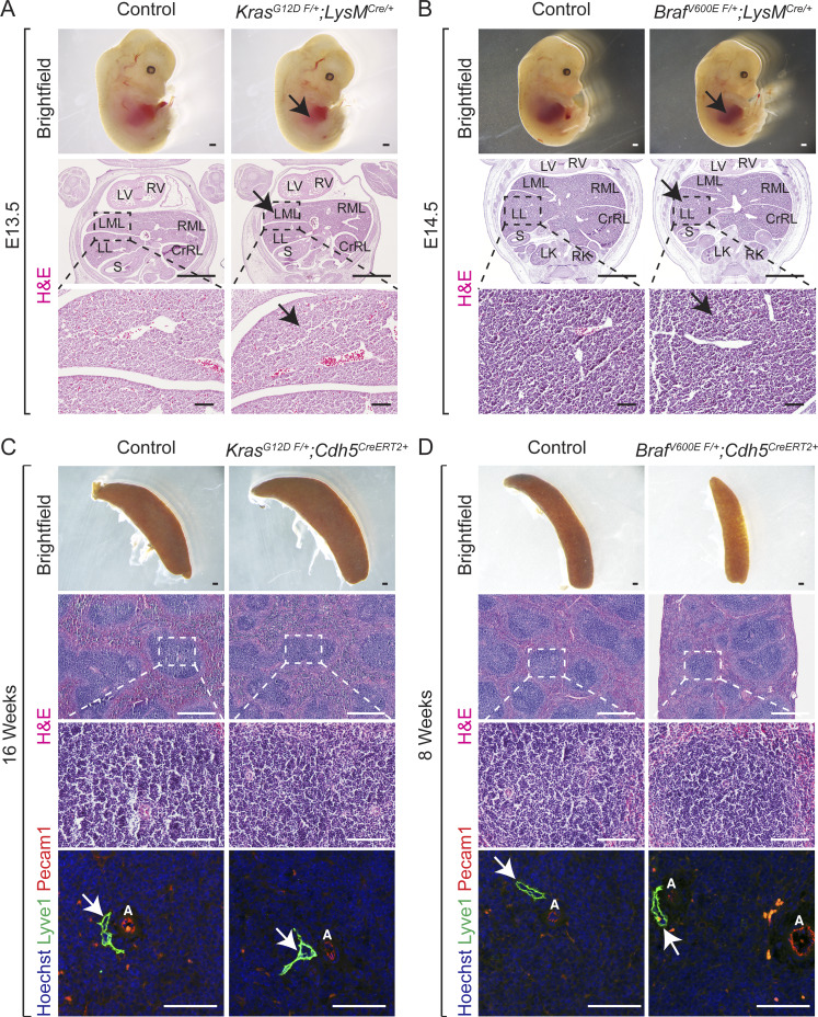
Figure S4.
Mice with KrasG12D or BrafV600E gain-of-function mutations within macrophages exhibit normal normal sinusoidal and hepatic development. (A and B) Dissected KrasG12D F/+; LysMCre (A) and BrafV600E F/+; LysMCre (B) embryos show normal liver size (black arrows). H&E-stained KrasG12D F/+; LysMCre (A) and BrafV600E F/+; LysMCre (B) liver frontal sections show normal sinusoidal capillaries (black arrows). n = 3. E, embryonic day. Scale bars: 500 µm (top row), 1,000 µm (middle row), and 100 µm (bottom row). Two independent experiments. (C and D) Mice with KrasG12D or BrafV600E gain-of-function mutations within endothelial cells exhibit normal normal spleen morphology and sinusoids. Spleen dissected from adult tamoxifen-treated KrasG12D F/+; Cdh5CreERT2 (C) and BrafV600E F/+; Cdh5CreERT2 (D) mice. H&E-stained KrasG12D F/+; Cdh5CreERT2 (C) and BrafV600E F/+; Cdh5CreERT2 (D) spleen sagittal sections show normal morphology. Lyve1 (green) and Pecam1 (red) coimmunofluorescent staining with Hoechst nuclear counterstain (blue) on KrasG12D F/+; Cdh5CreERT2 (C) and BrafV600E F/+; Cdh5CreERT2 (D) spleen sagittal sections. n = 3. Scale bars: 500 µm (first and second rows), 100 µm (third row), and 50 µm (fourth row). Two independent experiments. White arrows show normal Lyve1+ vessels. A, artery; CrRL, cranial right lobe; LK, left kidney; LL, left lobe; LML, left medial lobe; LV, left ventricle; RK, right kidney; RML, right medial lobe; RV, right ventricle; S, stomach.

