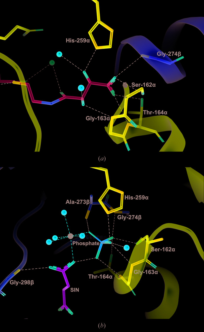
Figure 4.
Interactions between tartryl-CoA or phosphate and the phosphate-binding site of GTPSCS. (a) Tartryl-CoA and interacting residues are displayed as stick models. C atoms are shown in cyan for tartryl-CoA. (b) Phosphate, succinate and interacting residues of the protein in the structure of Mg2+-succinate-bound GTPSCS (PDB entry 5cae; Huang & Fraser, 2016 ▸) are drawn as stick models. C atoms are shown in green for succinate (abbreviated SIN). The magnesium ion is represented by a black sphere. The orientations are somewhat different to better show the substrates. For both structures, interacting residues of the α-subunit and β-subunit are shown as stick models with purple and yellow C atoms, respectively, water molecules are represented by red spheres and interactions are represented by dashed lines.

