Figure 5.

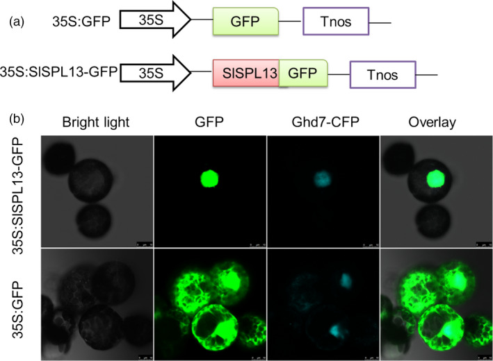
Subcellular localization of SPL13. (a) Schematic diagrams of the constructs used to determine subcellular localization. The SPL13 CDS without the stop codon was amplified by PCR and fused to the 5′ end of the open reading frame encoding GFP in pCAMBIA 1302. The expression of SPL13‐GFP was driven by the CaMV 35S. (b) Transient expression of 35S:SPL13‐GFP and 35S:GFP in tobacco (N. benthamiana) protoplasts. The nuclei were identified by co‐expressing the nuclear marker Ghd7‐CFP with both 35S:SPL13‐GFP and 35S:GFP. Fluorescence images were acquired using a confocal laser scanning microscope (Leica TCS SP2, MRC Centre for Regenerative Medicine, The University of Edinburgh, Edinburgh, UK) after incubating the protoplasts at 28 °C for 12–16 h. Representative micrographs are shown. Bars, 10 μm.
