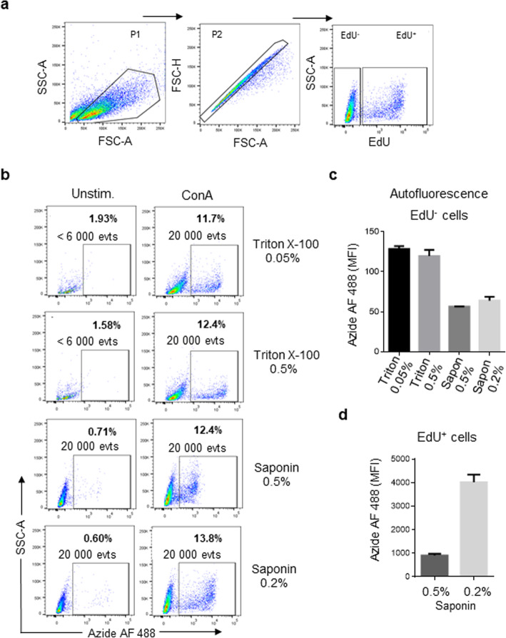
Fig. 2.
Effect of permeabilization reagents on the detection of EdU+ cells. Mononuclear splenocyte cells, cultured for 72 h in the presence or absence of 1 μg/ml ConA, were fixed and treated with different permeabilization reagents (saponin or Triton X-100). (a) Flow cytometry analysis for detecting EdU incorporation into cells. (b) Representing dot plots of the cells treated with different permeabilization reagents. (c) Comparison of median fluorescence intensity (MFI) in EdU− cells (autofluorescence) treated with saponin or Triton X-100. (d) Comparison of MFI of EdU+ cells treated with 0.5% or 0.2% saponin. Results are the mean ± standard deviation from of 2 independent experiments, performed in duplicate. In each experiment, the cells of one chicken were analyzed. Per sample, 20,000 events were acquired on a FACSMelody flow cytometer

