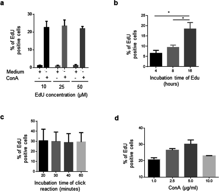
Fig. 3.
Click reaction conditions. Mononuclear cells isolated from the spleen were cultured in duplicate for 72 h in the presence or absence of 1 μg/ml ConA. The same flow cytometry analysis showed in Fig. 2a was followed. (a) Percentage of EdU+ cells after 4 h incubation with 10, 25, or 50 μM of EdU. The results are expressed as the mean ± standard deviation of 3 independent experiments. (b) Percentage of EdU+ cells incubated with 25 μM of EdU for increasing time. The results are expressed as the mean ± standard deviation of 3 independent experiments. (c) Percentage of EdU+ cells after incubating activated cells with click reaction components for increasing time. The results are expressed as the mean ± standard deviation of 3 independent experiments. (d) Percentage of EdU+ cells stimulated with increasing concentrations of 1 μg/ml ConA. EdU (25 μM) was added at 16 h before the end of the culture. The staining time with the click reaction solution was 20 min. The results are expressed as the mean ± standard deviation of 2 independent experiments. In each experiment, the cells of 1 chicken were analyzed. All values shown are percentage of singlet cells. Significant differences are indicated by * (p = 0.0286). Per sample, 30,000 events were acquired on a FACSMelody flow cytometer

