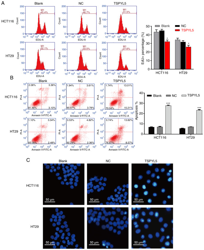
Figure 3.
Effects of TSPYL5 overexpression on the proliferation and apoptosis of CRC cells. HCT116 and HT29 cells were transfected with pcDNA3.1-TSPYL5 or empty pcDNA3.1, and subsequently divided into TSPYL5 and negative control (NC) groups, respectively. (A) EdU flow cytometry was used to analyze the percentage of EdU-positive cells. (B) Flow cytometric analysis of cell apoptosis rates using Annexin V-FITC/PI double labeling. Data represent the mean ± SD of results from three individual experiments; ***P<0.001 vs. NC. (C) Fluorescence assay of Hoechst 33342 staining. HCT116 and HT29 cells were stained with Hoechst 33342 staining solution and visualized under a fluorescence microscope. CRC, colorectal cancer; TSPYL5, testis-specific protein Y-encoded-like 5.

