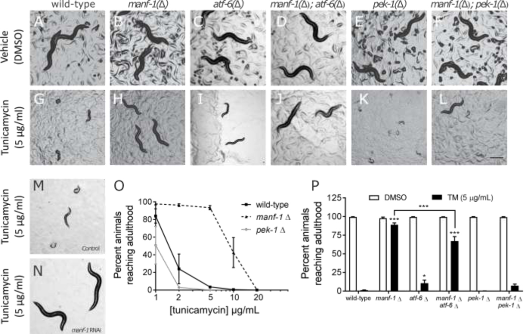
Figure 3. MANF-1 deficiency confers resistance to tunicamycin-mediated developmental arrest.
Worms harboring mutations (indicated by “Δ”) in manf-1 (B), atf-6 (C), and pek-1 (E) have developmental rates similar to wildtype animals (A) when hatched under normal conditions. Similar results were obtained when the manf-1 mutation is paired with either the atf-6 or the pek-1 mutant (D, F). Wild-type (G) and atf-6 mutant (I) larva from embryos hatched in the presence of tunicamycin exhibit delayed or arrested development, while pek-1 mutants die (K). Manf-1 mutant larvae appear unaffected by tunicamycin (H). The tunicamycin-resistant phenotype was also observed in the manf-1; atf-6 double mutant (J), although not to the same extent. The manf-1; pek-1 double mutant was only slightly more resistant to tunicamycin compared to the pek-1 single mutant, and not statistically significant (L). RNAi knockdown of manf-1 also confers resistance to tunicamycin compared to control RNAi (M,N). Dose response to tunicamycin showing the effects on the larval development up to adulthood (O). The percentage of animals (n=100 per condition) reaching adulthood in the presence of tunicamycin (5 μg/mL) for wild-type, manf-1, atf-6, pek-1 as single mutants, and in combination (P). Data shown are the average of four biological replicates. Asterisks represent statistical significance in a two-way ANOVA (dose, strain) compared to the tunicamycin response in wild-type in a Bonferroni post-test unless otherwise indicated.

