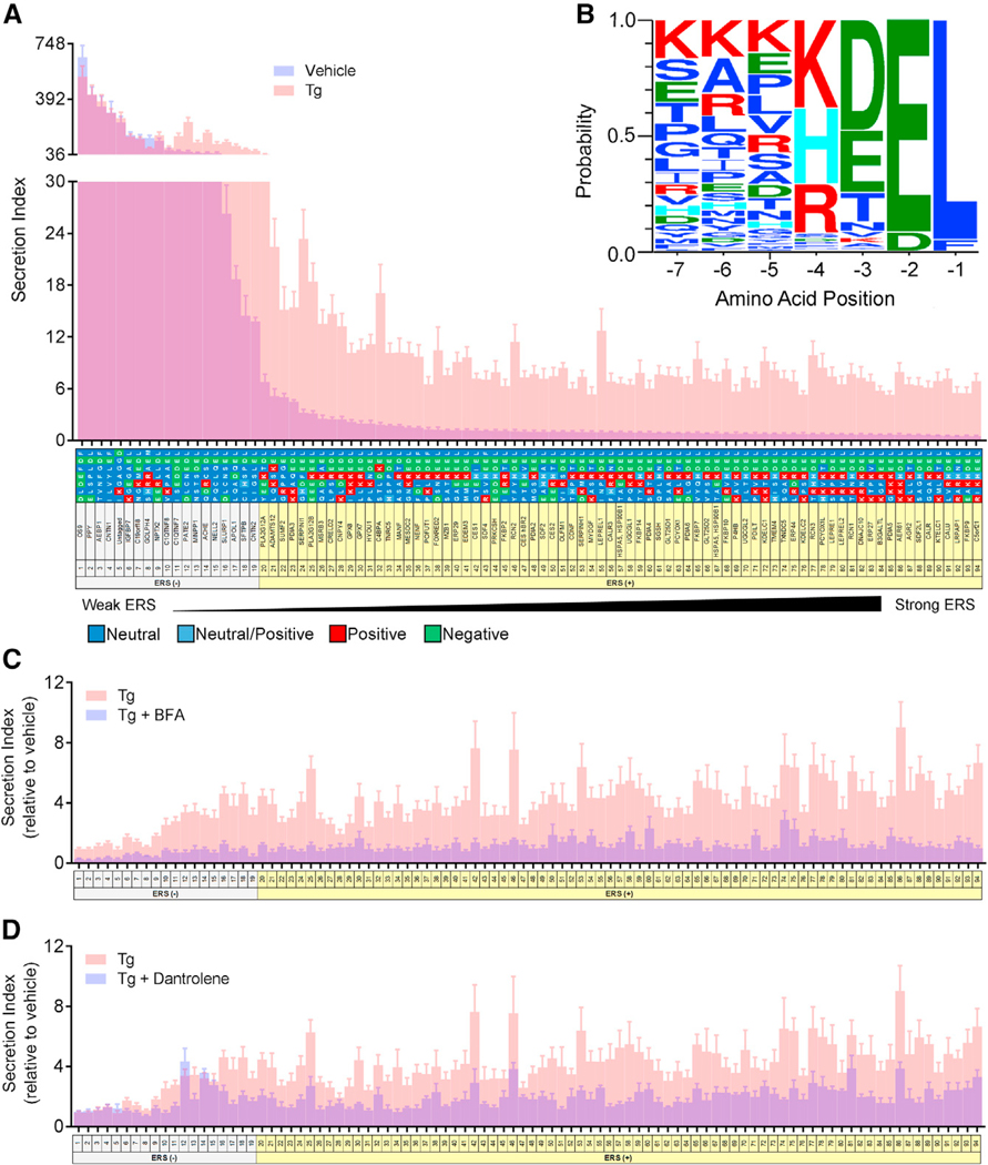
Figure 1. Characterizing the Secretion of ER Resident Proteins with ERSs.
(A) Secretion of GLuc fusion proteins from transiently transfected SH-SY5Y cells under basal conditions (blue) and after 200 nM Tg for 24 hr (pink) (n = 9). Overlap of the two groups is purple. Secretion index refers to the ratio of extracellular to intracellular GLuc.
(B) Sequence logo produced by WebLogo 3.4 forproteins designated as ERS (+).
(C and D) Fold change in secretion of GLuc fusion protein from transiently transfected SH-SY5Y with (blue) or without (pink) (C) 500 nM brefeldin A (BFA) or (D) 50 μM dantrolene prior to an 8 hr exposure to 200 nM Tg (n = 6 for dantrolene, n = 12 for Tg).
See also Figures S1 and S2.

