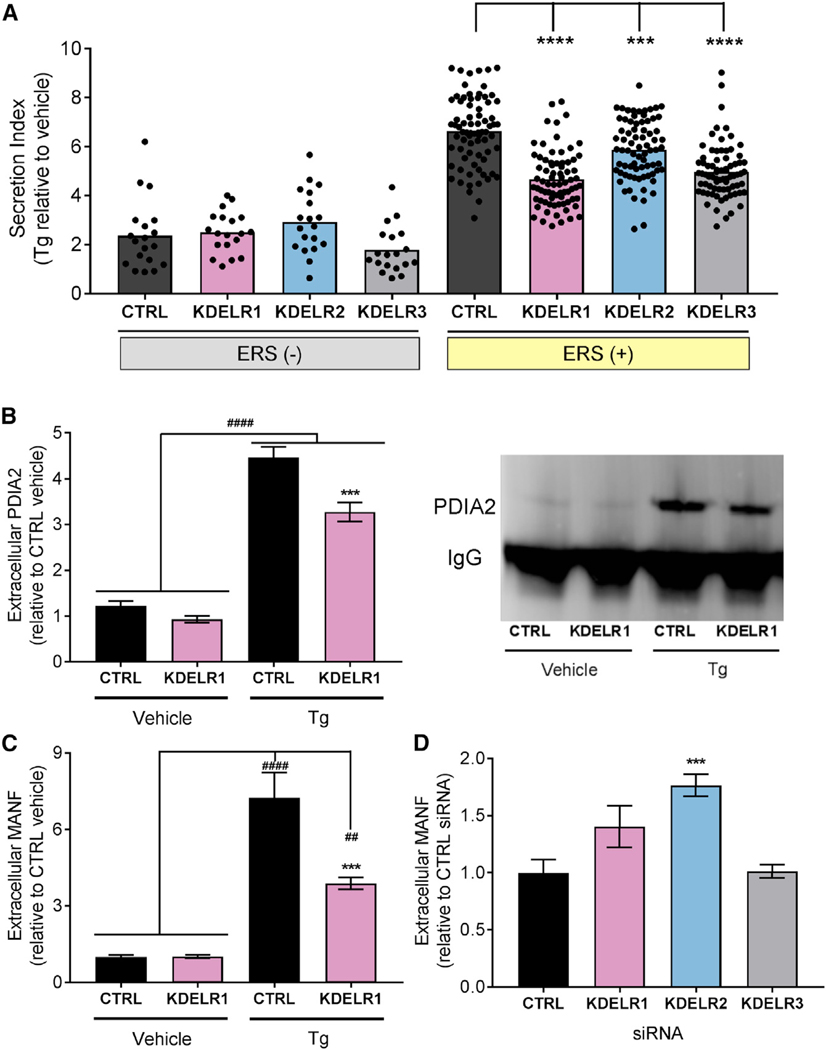
Figure 4. KDEL Receptors Regulate the Secretion of ERS-Containing Proteins.
(A) Average fold change in secretion of ERS (+) and ERS (−) constructs from transiently transfected SH-SY5Y cells overexpressing KDEL receptors following treatment with 200 nM Tg for 24 hr (n = 4; two-way ANOVA with Dunnett’s multiple comparisons test; p < 0.0001 for ERS (+) versus ERS (−), ***p < 0.001 and ****p < 0.0001 for control versus KDEL receptor). Each dot represents a unique GLuc-ERS.
(B and C) Fold change in (B) immunoprecipitated PDIA2 (representative blot is shown to the right) or (C) MANF in media from SH-SY5Y cells overexpressing KDELR1 or control and treated with 200 nM Tg or vehicle for 24 hr (n = 9; **p < 0.01, ####p < 0.0001 Tg versus vehicle, ***p < 0.001 KDELR1 Tg versus CTRL Tg). (C) Extracellular MANF levels in the media of SH-SY5Y cells transfected with KDEL receptor siRNAs (n = 6; one-way ANOVA and Dunnett’s test; ***p < 0.001 versus CTRL).
See also Figures S4 and S5.

