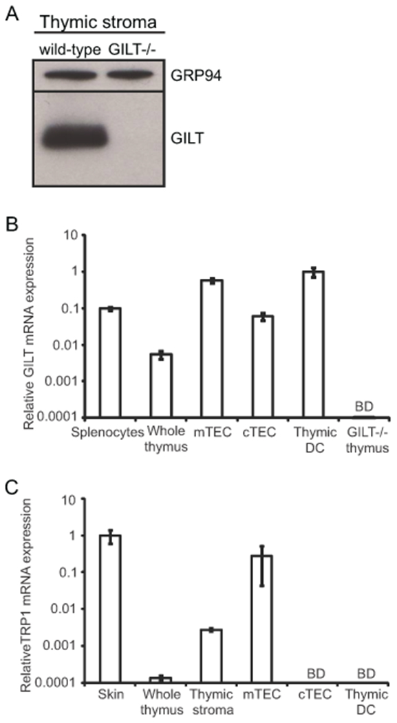
Figure 2. GILT expression is enriched in mTECs and thymic DCs, and TRP1 expression is limited to mTECs.

(A) Immunoblot analysis demonstrating GILT expression in total thymic stromal cells isolated by density gradient centrifugation. GRP94 was used as a loading control. (B) Thymic stromal cells were FACS-sorted to obtain purified populations of CD45+CD11c+ dendritic cells (DCs), CD45-EpCAM+Ly51-UEA-1+ medullary thymic epithelial cells (mTECs), and CD45-EpCAM+Ly51+UEA-1− cortical thymic epithelial cells (cTECs). GILT transcript levels assessed by quantitative PCR of splenocytes (positive control), whole thymus, mTECs, cTECs, thymic DCs, and GILT−/− thymus (negative control). Data were normalized to cyclophilin and shown relative to thymic DCs. (C) TRP1 transcript levels assessed by quantitative PCR of skin (positive control), whole thymus, total thymic stromal cells, mTECs, cTECs, and thymic DCs. Data were normalized to cyclophilin and shown relative to skin. Unless otherwise indicated, cells were obtained from wild-type mice. BD, below the limit of detection.
