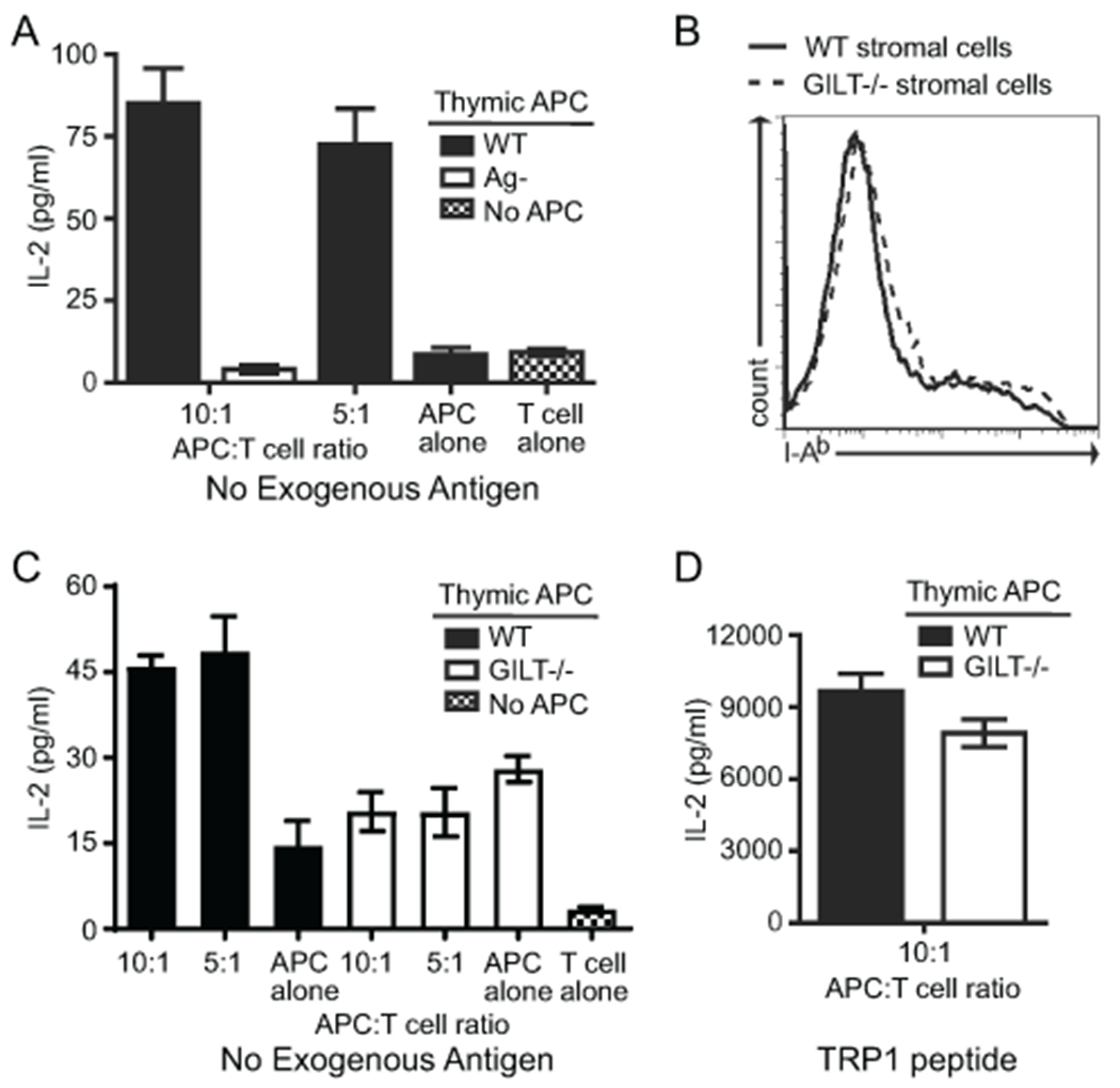
Figure 3. GILT facilitates MHC class II-restricted presentation of endogenous TRP1 by pooled thymic APCs.

(A) Pooled thymic stromal cells isolated by density gradient centrifugation were co-cultured with primary, naive TRP1-specific T cells without additional antigen. Thymic APCs from TRP1-deficient (Ag−) mice served as a negative control. IL-2 production, as assessed by ELISA, was used as a measure of antigen presentation. Comparison of wild-type APCs (both 10:1 and 5:1) with each of the negative controls (thymic APCs from TRP1-deficient (Ag−) mice, wild-type APCs alone, and T cells alone) by one way ANOVA followed by Tukey’s multiple comparison test revealed p < 0.001 for each. (B) I-Ab expression on thymic stromal APCs from WT and GILT−/− mice. (C) Pooled thymic stromal cells from WT and GILT−/− mice were co-cultured with naive TRP1-specific T cells without additional antigen. Comparison of wild-type APCs 10:1 and 5:1 with wild-type APCs alone and T cells alone revealed p < 0.001 for each; no significant differences between GILT−/− APCs 10:1 and 5:1 with GILT−/− APCs alone and T cells alone. (D) Pooled thymic stromal cells from WT and GILT−/− mice were co-cultured with naive TRP1-specific T cells with TRP1 peptide (10 μg/ml) (positive control). No significant difference identified by an unpaired t test. Bars and error bars represent the mean ± SEM of triplicates in one experiment using the number of mice required to obtain sufficient cells. The data are representative of three independent experiments.
