
FIGURE 5.
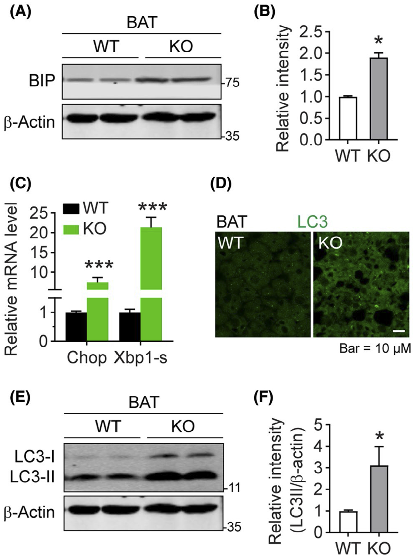
Deficiency of DRP1 triggers ER stress and autophagy in adipose tissue. A, Western blotting analysis of BIP (ER stress marker protein) and β-Actin (as loading control) protein levels in the BAT from Adipo-Drp1flx/flx mice and their littermate controls after cold exposure. B, Quantification of relative band intensity for BIP (normalized to β-Actin) in (A) (n = 6 per group, the sample loaded in each lane contains pooled samples from three mice, representative of three trials. The data are represented as mean ± SEM, Student’s t test, *P < .05). C, Q-PCR analysis of the mRNA levels for Chop and Splice-Xbp1s (Xbp1-s) (ER stress related genes) in the BAT from Adipo-Drp1flx/flx mice and their littermate controls after cold exposure (n = 5 per group, data are represented as mean ± SEM, Student’s t test, ***P < .001). D, IF staining with anti-LC3 antibody in the BAT from Adipo-Drp1flx/flx mice and their littermate controls after cold exposure (Representative of three trials are shown). E, Western blotting analysis of LC3-I/II and β-Actin protein levels in the BAT of Adipo-Drp1flx/flx mice and their littermate controls after cold exposure. F, Quantification of relative band intensity for LC3-II (normalized to β-Actin) in (E) (n = 6 per group, each lane contains pooled samples from three mice, representative of three trials. The data are represented as mean ± SEM, Student’s t test, *P < .05)