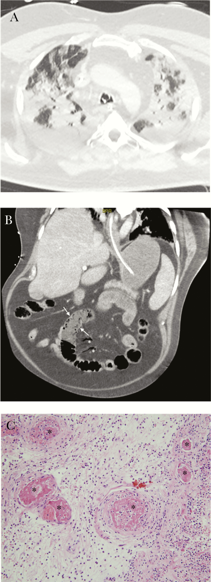
Figure 1.
Lungs and gut are shown. (a) Initial chest computed tomography (CT) scan: extensive bilateral ground-glass opacities and alveolar consolidation, with peripheral and subpleural predominance, in keeping with a severe coronavirus disease 2019 (COVID-19). No evidence of pulmonary embolism was present at that time. (b) Abdominal CT: bowel wall thickening with severe hypoenhancement (see Figure 2b, arrows) and significant mesenteric intravenous air (see Figure 2b, stars), suggestive of mesenteric ischemia. (c) Small intestine histology: thrombosed small blood vessels in submucosal bowel at higher magnification (stars, hematoxylin and eosin stain ×200).

