Figure 4.
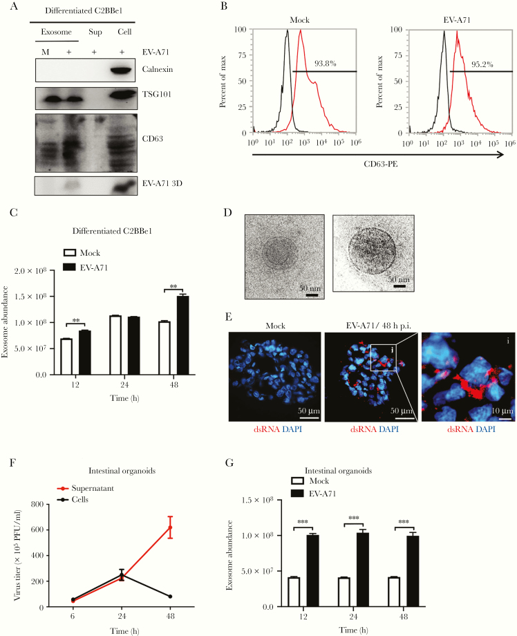
Enterovirus A71 (EV-A71) increases the release of exosomes from differentiated C2BBe1 cells and human intestinal organoids. (A) Differentiated C2BBe1 cells were infected with EV-A71 at a multiplicity of infection of 10 for 24 hours. Exosomes purified from mock- and EV-A71-infected samples were subjected from total protein isolation. Immunoblotting analysis was performed using antibodies specific for CD63, calnexin, TSG101, and EV-A71 3D. Total protein extracted from supernatants and cells collected from infected samples were used as control. (B) Flow cytometric results showing the percentage of CD63-positive vesicles released from mock- and EV-A71-infected differentiated C2BBe1 cells. (C) FluoroCet Exosome quantitation kit was performed to quantify the exosomes extracted from differentiated C2BBe1 cells that were mock and EV-A71 infected. (D) Cryoelectron microscopy images of the exosomes purified from the differentiated C2BBe1 cells infected with EV-A71. (E) Intestinal organoids were infected with EV-A71 for 48 hours. Immunofluorescence staining was performed using antibodies specific for double-stranded ribonucleic acid (dsRNA). (scale bar, 50 μm/10 μm). (F) Supernatants and infected cells were collected from infected human organoids, and viral titers were quantified by plaque-forming assay. (G) FluoroCet Exosome quantitation kit was performed to quantify the exosomes extracted from mock- and EV-A71-infected intestinal organoids. The data are presented as the means + standard deviation. Significant differences between 2 groups were determined by Student’s t test (**, P < .01; ***, P < .001). DAPI, 4’,6-diamidino-2-phenylindole; h p.i., hours postinfection; PFU, plaque-forming units.

