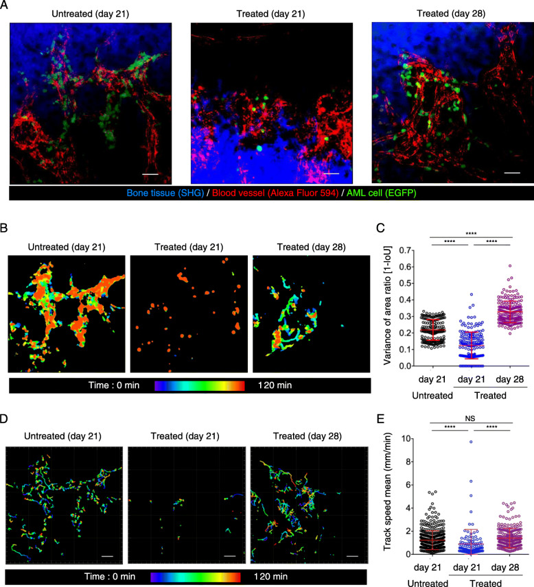
Fig. 1.
Dynamics of AML cells in bones during chemotherapy. a Representative intravital two-photon maximum-intensity projection (MIP) skull images at 21 and 28 days after AML cell transplantation. Images from untreated AML-transplanted mice at day 21, treated AML-transplanted mice at day 21 and treated AML-transplanted mice at day 28 are shown. Green, GFP-expressing AML cells; red, blood vessels (Alexa Fluor 594); blue, bone tissues (second harmonic generation; SHG). Scale bar, 50 μm. See also Video 2. b Representative images showing AML cell area. Scale bar, 50 μm. c Scatter plots showing displacement area ratio. Data are presented as mean ± SD; n = 3 mice per group; ****P < 0.0001 (one-way ANOVA). d Representative images of migrating AML cell trajectories. Scale bar, 50 μm. e Scatter plots showing the mean track speed of all cells analyzed in (d). Data from three mice per group from independent experiments are shown. Untreated (21 days), n = 461; treated (21 days), n = 108; treated (28 days), n = 243. Data are presented as mean ± SD. ****P < 0.0001; NS, not significant (one-way ANOVA)

