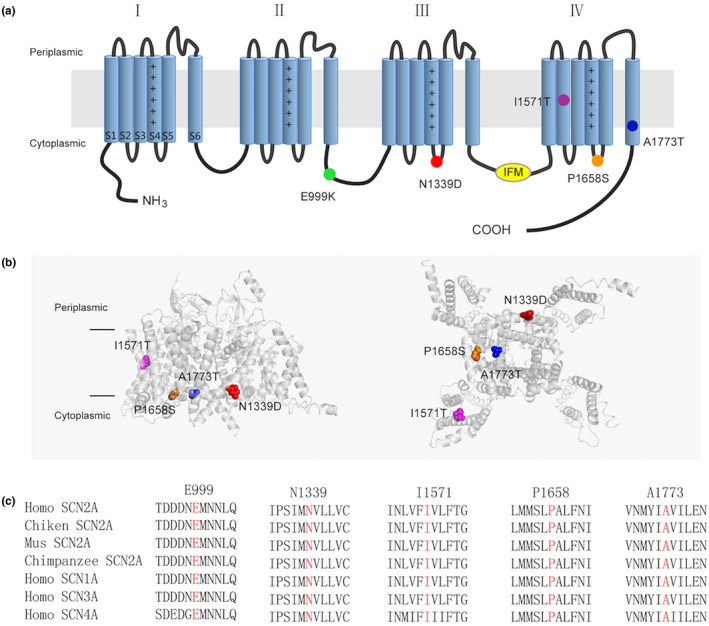
FIGURE 1.

Locations of Epilepsy‐associated Nav1.2 mutations. (a) Topology diagram of the human Nav1.2 channel α subunit. The α subunit consists of four homologous domains (I–IV), each of which contains six transmembrane regions (S1–S6). Plus signs in S4 represent the positively charged voltage sensor (containing a number of arginines or lysines). Segments S1−S3 and S4 form the voltage‐sensing domain. S5–S6 in conjunction with their extracellular linker constitute the channel pore. The intracellular loop connecting III/S6 and IV/S1 contains the isoleucine, phenylalanine, and methionine (IFM) domain involved in channel inactivation (yellow). The locations of the mutated residues described in the present report are shown as circles in different colors. (b) Structure of the human Nav1.2 α subunit (PDB:6J8E). The left is the side view and the right is the bottom view. The mutations are shown as CPK style in different colors. The p.E999K mutation is located in the loop between II and III, which are not resolved in the original structure, so it cannot be exhibited. (c) Protein alignment showing that the affected residues (highlighted in red) are conserved cross in human Nav homologous and various species orthologs
