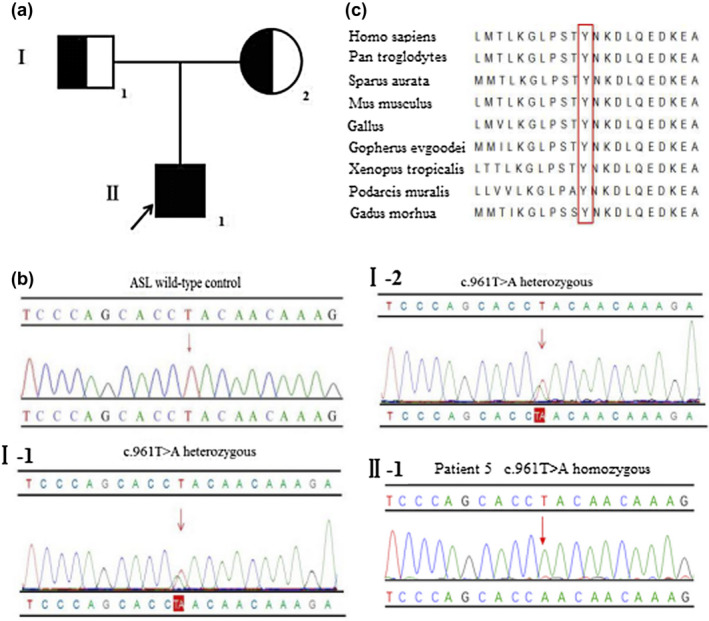
Figure 3.

(a) Pedigree of patient 5. The arrow denotes the proband. (b) Sanger sequencing analysis of the ASL, respectively, identified the mutation c.961T>A (p.Tyr321Asn) in exon 13 (Ⅰ‐1) heterozygous in his father, (Ⅰ‐2) heterozygous in his mother, and (Ⅱ‐1) homozygous in patient 5 (the variant is indicated by a red arrow). (c) Multiple sequence alignment using Clustal X. The tyrosine residue at position 321 (highlighted by a red box) is highly conserved among different species
