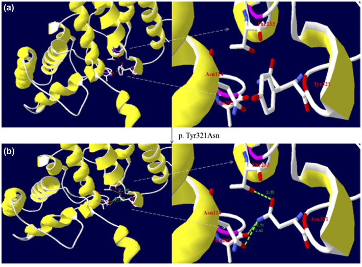
Figure 4.

Three‐dimensional modeling structure analysis of wild‐type and mutant products of ASL. Green dashed lines represent hydrogen bonds and the green number shows the hydrogen bonds distances. (a) A segment of the ASL structure showing Tyr321 has a side chain of benzene and it has no hydrogen bonds with the adjacent domain. (b) A segment of the ASL structure showing that Asn321 has a side chain without benzene and that its backbone makes new hydrogen bonds with the side chain of Asn114, the backbone of Ala203, and the side chain of Thr233. The disappearance of the bulky and rigid benzene side chain and new hydrogen bonds may induce a distortion of the ASL protein structure. (The color in this figure is that selected by the Secondary Structure Succession of Swiss‐PdbViewer 4.10.)
