. 2020 Jul 2:202352. doi: 10.1148/radiol.2020202352

Figure 3c:

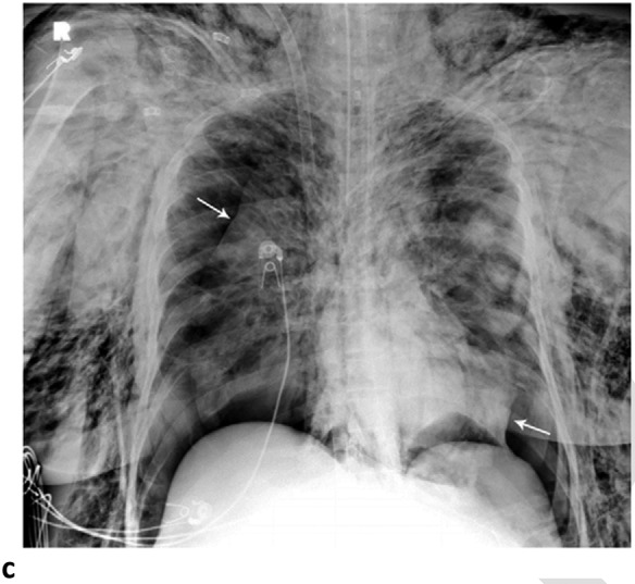
Pneumomediastinum and Bilateral Pneumothoraces, Separated by 7 days. 20-year-old woman intubated 5 days after admission. (a) Frontal chest radiograph depicts moderate pneumomediastinum (black arrows) and subcutaneous emphysema. (b) Frontal radiograph 3 days later demonstrates resolution of pneumomediastinum, and persistent mild subcutaneous emphysema. The superior and inferior extracorporeal membrane oxygenation catheters (arrowheads) were placed the preceding day. (c). Four days later, she developed large bilateral pneumothoraces (white arrows), and extensive subcutaneous emphysema.

Pneumomediastinum and Bilateral Pneumothoraces, Separated by 7 days. 20-year-old woman intubated 5 days after admission. (a) Frontal chest radiograph depicts moderate pneumomediastinum (black arrows) and subcutaneous emphysema. (b) Frontal radiograph 3 days later demonstrates resolution of pneumomediastinum, and persistent mild subcutaneous emphysema. The superior and inferior extracorporeal membrane oxygenation catheters (arrowheads) were placed the preceding day. (c). Four days later, she developed large bilateral pneumothoraces (white arrows), and extensive subcutaneous emphysema.