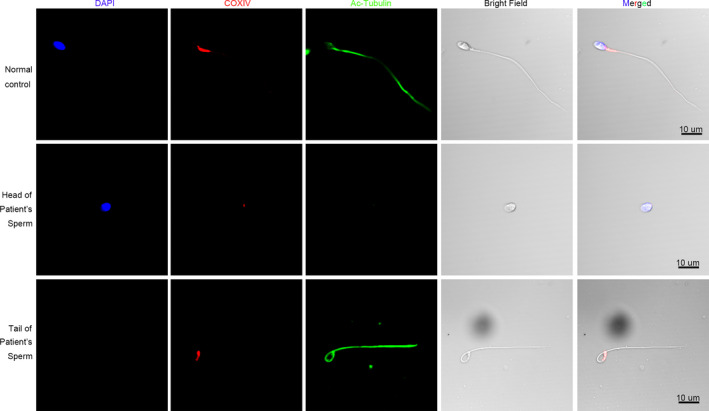
FIGURE 5.

COXIV protein expression in the patient and normal control. COXIV (Red) protein localization was determined by immunofluorescence assay. The nuclei were stained with DAPI (Blue). Multiple images were taken and the representative ones were presented. Scale bar: 10 μm
