. 2020 Jun 5;40(4):E8–E15. doi: 10.1148/rg.2020200097

Figure 1.

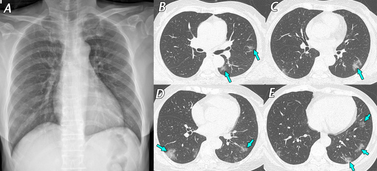
Mild cough and dizziness 1 day before visiting a screening clinic in a 62-year-old woman with contact history with a confirmed SARS-CoV-2–infected patient 4 days earlier. RT-PCR test results were positive for SARS-CoV-2, and she was transferred to our hospital and admitted to a containment zone. Initial chest CT findings (not shown) were normal. A, Posteroanterior (PA) chest radiograph obtained 9 days after initial symptom onset shows no definite abnormality. B–E, Follow-up axial chest CT images obtained on the same day as the chest radiograph show multifocal ground-glass opacities (arrows), predominantly located in the peripheral areas of both lungs. After 13 days of conservative management, her respiratory symptoms ameliorated, and a negative RT-PCR test result for SARS-CoV-2 was obtained. RT-PCR was repeated twice to confirm this negative result.

Mild cough and dizziness 1 day before visiting a screening clinic in a 62-year-old woman with contact history with a confirmed SARS-CoV-2–infected patient 4 days earlier. RT-PCR test results were positive for SARS-CoV-2, and she was transferred to our hospital and admitted to a containment zone. Initial chest CT findings (not shown) were normal. A, Posteroanterior (PA) chest radiograph obtained 9 days after initial symptom onset shows no definite abnormality. B–E, Follow-up axial chest CT images obtained on the same day as the chest radiograph show multifocal ground-glass opacities (arrows), predominantly located in the peripheral areas of both lungs. After 13 days of conservative management, her respiratory symptoms ameliorated, and a negative RT-PCR test result for SARS-CoV-2 was obtained. RT-PCR was repeated twice to confirm this negative result.