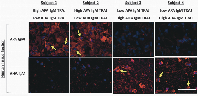
FIG. 10.
Anti-pituitary antibody (APA) and anti-hypothalamic antibody (AHA) immunoglobulin (Ig)M fluorescence immunohistochemistry (IHC) staining with selected traumatic brain injury (TBI) participants. Human cavaderic pituitary and hypothalamic tissue was stained with subacute-chronic (1 month) serum samples from four men (subjects 1–4) with moderate to severe TBI. Each serum sample was exposed to human pituitary and hypothalamic tissue sections and then developed with fluorophore Alexa 555-conjugated anti-human IgM (images shown from top to bottom in each column). Yellow arrows point to strongly staining cells. White bar in lower right corner represents 50 μm. Individual subject's membership in APA and AHA IgM TRAJ group (high, low) are shown on top and bottom, respectively, of its IHC images.

