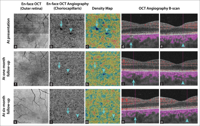
Figure 2.
En-face swept-source optical coherence tomography (SS-OCT) and SS-OCT angiography (SS-OCTA) at presentation in the same patient of Figure 1 (a-e): Structural en-face SS-OCT (3 mm × 3 mm scans) taken at the level of the outer retina revealing hyporeflective areas corresponding to the ellipsoid zone disruptions. En-face SS-OCTA at the level of choriocapillaris showing two small areas of hypointense flow deficit, which are highlighted by respectively an arrow and an arrowhead, with decreased flow signal in the choriocapillaris on respective SS-OCTA B-scans. No evidence of any shadow graphic or projection artifacts is appreciable. En-face SS-OCT and SS-OCTA, at 1-month follow-up (f-j): En-face SS-OCTA, 1 month after the initial presentation, showing the resolution of the choriocapillaris hypointense areas with the restitution of flow signals in the choriocapillaris on SS-OCTA B-scan, and the persistence of a diffuse hyporeflective area on en-face SS-OCT at the level of the outer retina. En-face SS-OCT and SS-OCTA, at 6-month follow-up (k-o): En-face SS-OCT at the level of the outer retina and SS-OCTA at the level of the choriocapillaris showing quite normal findings

