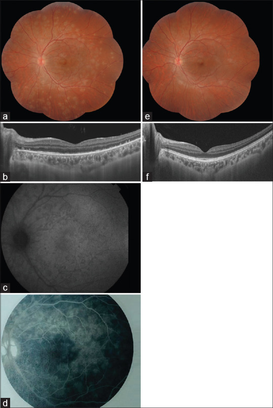
Figure 3.

Multimodal imaging of a 24-year-old patient during the acute phase of multiple evanescent white dots syndrome. (a) Composite color fundus photograph of the left eye showing multiple, deep, white dots in the posterior pole and mid-periphery and macular granularity. (b) Structural swept-source optical coherence tomography (SS-OCT) demonstrating disruption of the ellipsoid zone with small accumulations of hyperreflective material resting over the retinal pigment epithelium and extending toward the outer nuclear layer. (c) Near-infrared fundus autofluorescence showing hypoautofluorescent spots. (d) Fluorescein angiography during late-frames showing the multifocal hyperfluorescence at the posterior pole associated with optic disc leakage. (e) Composite color fundus photograph of the left eye and (f) SS-OCT, 1 year after the first presentation, showing the complete resolution of abnormal findings
