Figure 4.
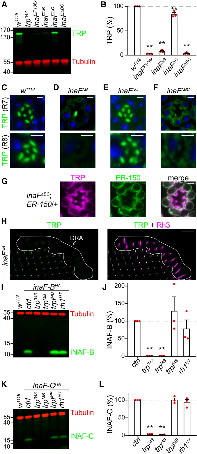
INAF-B/INAF-C and TRP are mutually required for expression in photoreceptor cells. (A, I, and K) Head extracts were prepared from the indicated flies, 0.5 head equivalents/lane were fractionated by SDS-PAGE, and Western blots were probed with mouse anti-tubulin (loading control) and the following antibodies: (A) rabbit anti-TRP, (I) rabbit anti-HA (recognizes INAF-B::HA), and (K) rabbit anti-HA (recognizes INAF-C::HA). The anti-HA signals reflected the endogenous levels of INAF-B and INAF-C since the extracts were prepared from flies with HA-tags knocked into the endogenous inaF-B and inaF-C genes. Protein size markers are indicated (kDa). (B, J, and L) Relative levels of the following proteins in heads of the indicated flies, based on Western blot analyses shown to the left: (B) TRP, (J) INAF-B, and (L) INAF-C; n = 3. Error bars represent SEMs; **P < 0.01 (one-way ANOVA with Holm–Sidak post hoc analyses). (C–F) Optical sections from single-ommatidia from the compound eyes of w1118 flies and the indicated inaF mutants, stained with rabbit anti-TRP (green) and a nuclear counterstain, To-PRO3 (blue). Top: R7 layer; bottom: R8 layer; bar, 3 μm. (G) Optical sections from single-ommatidia (R7 layer) from the compound eyes of inaFΔBC;ER-150/+ flies, stained with rabbit anti-TRP (magenta) and chicken anti-GFP (green, recognizes ER-150). ER-150 is an ER-localized GCaMP, which was expressed under control of the ninaE promoter (Liu et al. 2020); bar, 3 μm. (H) Multiple ommatidia (R7 layer) from inaFΔB flies around the DRA, were costained with rabbit anti-TRP (green) and mouse anti-Rh3 (magenta). The left panel shows staining with anti-TRP, while the right panel shows anti-TRP and anti-Rh3 staining; bar, 20 μm.

