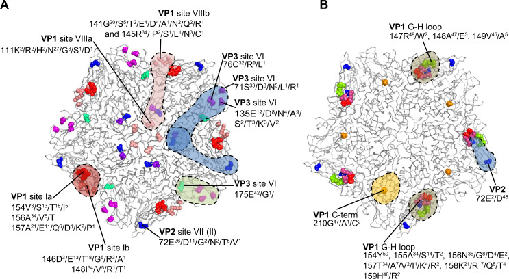
Figure 2.
The antigenic structure of foot-and-mouth disease SAT1 (A) and SAT2 (B) viruses are depicted.
Notes: The amino acid substitutions observed in SAT1 and SAT2 monoclonal antibody-resistant (MAR) mutants are shown as spheres on the grey ribbon backbone of the pentamer (five copies of VP1, VP2, and VP3) unit, and the potential antibody footprints are stipulated. The variation in amino acid residues at each antigenic residue position, in a complete capsid protein alignment of 43 SAT1 and 50 SAT2 viruses available on the genetic sequence database (GenBank) are indicated. The viruses in the alignments include isolates from South Africa, Zimbabwe, Mozambique, Namibia, Botswana, Zambia, Angola, Malawi, Tanzania, Kenya, Uganda, Rwanda, Zaire, Nigeria, Senegal, Ghana, Eritrea, Saudi Arabia, and Sudan. Adapted from Grazioli S, Moretti M, Barbieri I, Crosatti M, Brocchi E. Use of monoclonal antibodies to identify and map new antigenic determinants involved in neutralization of FMD viruses type SAT 1 and SAT 2 In: European Commission for the Control of Foot-and-Mouth Disease: International control of Foot-and-Mouth disease: Tools, Trends and perspectives. Paphos, Cyprus: Food and Agriculture Organization; 2006:287–297.85
Abbreviation: SAT, Southern African Territories.

