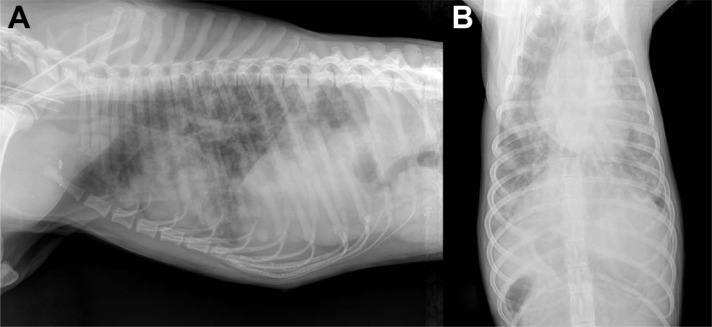
Figure 1.
Laterolateral (A) and dorsoventral (B) view of thorax of an 18-month-old dog: moderate interstitial pattern mainly involving the caudodorsal lung fields and mild alveolar pattern with isolated fluffy infiltrates and a subtle right-sided cardiac enlargement (Courtesy of Dr Paolo Crisi, Teaching Veterinary Hospital, University of Teramo, Italy).

