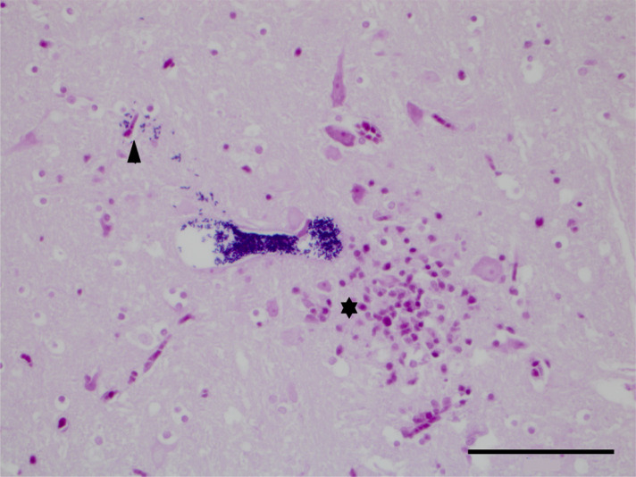
Figure 1.
Brainstem: a parasitophorous vacuole with distinct Gram-positive 1.5 × 2.5 µm spores. A few single Gram-positive spores can be seen escaping from the vacuole into the adjacent neuropil (arrowhead). There are increased numbers of microglial cells and astrocytes (gliosis) and likely a few lymphocytes, all of which stain pink, and a few viable neurons (⋆). (Gram’s stain, 400× magnification, bar 50 µm).

