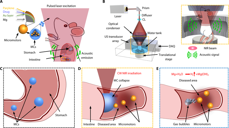
Fig. 1. Schematic of PAMR in vivo.
(A) Schematic of the PAMR in the GI tract. The MCs are administered into the mouse. NIR illumination facilitates the real-time photoacoustic imaging of the MCs, and subsequently triggers the propulsion of the micromotors in targeted areas of the GI tract. (B) Schematic of PACT of the MCs in the GI tract in vivo. The mouse was kept in the water tank surrounded by an elevationally focused ultrasound transducer array. NIR side-illumination onto the mouse generates PA signals, which are subsequently received by the transducer array. Inset figure: Enlarged view of the cyan dashed box region, illustrating the confocal design of light delivery and PA detection. MC, micromotor capsule; US, ultrasound; CL, conical lens; DAQ, data acquisition system; NIR, near infrared. (C) Enteric coating prevents the decomposition of MCs in the stomach. (D) External continuous-wave (CW) NIR irradiation induces the phase transition and subsequent collapse of the MCs on demand in the targeted areas and activates the movement of the micromotors upon unwrapping from the capsule. (E) Active propulsion of the micromotors promotes the retention and cargo delivery efficiency in intestines.

