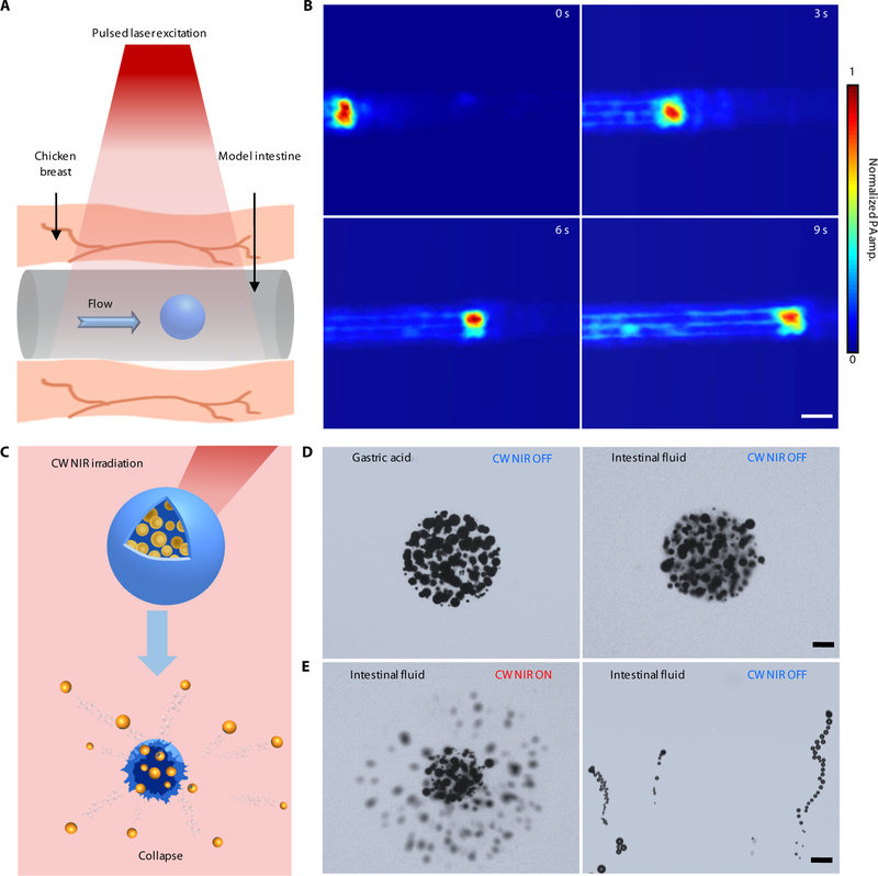
Fig. 3. Characterization of the dynamics of the PAMR.
(A and B) Schematic (A) and time-lapse PACT images in deep tissues (B) illustrating the migration of an MC in the model intestine. Scale bar, 500 μm. The thickness of the tissue above the MC is 10 mm. (C–E) Schematic (C) and time-lapse microscopic images (D and E) showing the stability of the MCs in gastric acid and intestinal fluid (D) without CW NIR irradiation, and the use of CW NIR irradiation to trigger the collapse of an MC and the activation of the micromotors (E). Scale bars in D and E, 50 μm.

