
Fig. 1.
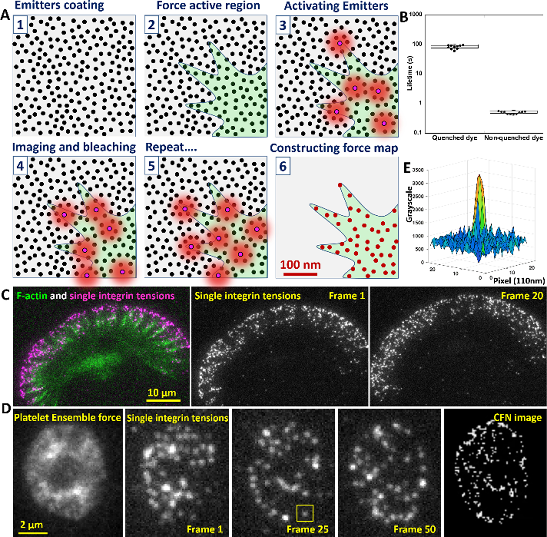
Principle of CFN and calibration of force-activatable emitter. (A) Emitters are coated on a surface at >2000/μm2 (step 1) to provide sufficient sampling density for cellular force (step 2). Emitters are sparsely switched on by local integrin tensions (step 3), imaged by a TIRFM and switched off by photobleaching (step 4). Multiple imaging cycles (step 5) and single molecule localization reconstructs a force map beyond the diffraction limit (Step 6). (B) The life time of Cy5 is extended by 120-fold in the proximity of a BHQ2 quencher, so that emitters prior to activation endure the switch-off process by photobleaching. (C) Demonstration of time-lapse imaging of integrin molecular tensions in a migrating keratocyte (Movie S1). (D) Ensemble force imaging and time-lapse imaging of integrin molecular tensions in a stationary platelet (Movie S3). (E) The grayscale map of an activated emitter (yellow squared region in Fig. 1D).