. 2020 Jun 5;40(4):1003–1019. doi: 10.1148/rg.2020190180

Figure 16b.

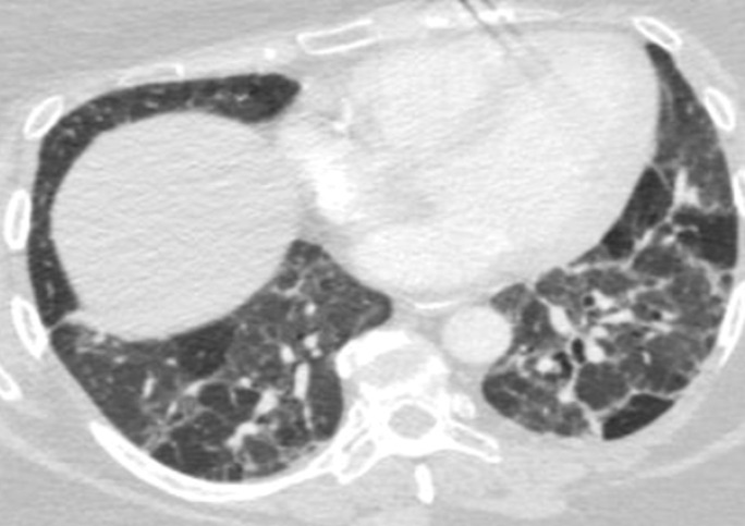
Variable CT imaging appearance of hypersensitivity pneumonitis in three patients. (a) Axial CT image in a 35-year-old man shows multiple centrilobular nodules. (b) Axial CT image in a 56-year-old woman shows mixed ground-glass opacity and mosaic attenuation interspersed with normal lung parenchyma (head cheese sign). (c, d) Axial CT images in a 46-year-old woman with a long history of using a hot tub shows areas of ground-glass attenuation predominantly in the left upper lobe (white oval in c). Evidence of air trapping appears on the expiratory image (d).

Variable CT imaging appearance of hypersensitivity pneumonitis in three patients. (a) Axial CT image in a 35-year-old man shows multiple centrilobular nodules. (b) Axial CT image in a 56-year-old woman shows mixed ground-glass opacity and mosaic attenuation interspersed with normal lung parenchyma (head cheese sign). (c, d) Axial CT images in a 46-year-old woman with a long history of using a hot tub shows areas of ground-glass attenuation predominantly in the left upper lobe (white oval in c). Evidence of air trapping appears on the expiratory image (d).