. 2020 Jun 5;40(4):1003–1019. doi: 10.1148/rg.2020190180

Figure 17b.

Progressive massive fibrosis in a 61-year-old man. Frontal chest radiograph (a) shows large bilateral upper lobe–predominant masses with irregular margins (arrows) and upper lobe volume loss, indicated by a juxtaphrenic peak (arrowhead). (b) Axial CT image shows bilateral large calcified conglomerate masses with adjacent fibrosis and calcified mediastinal lymph nodes.

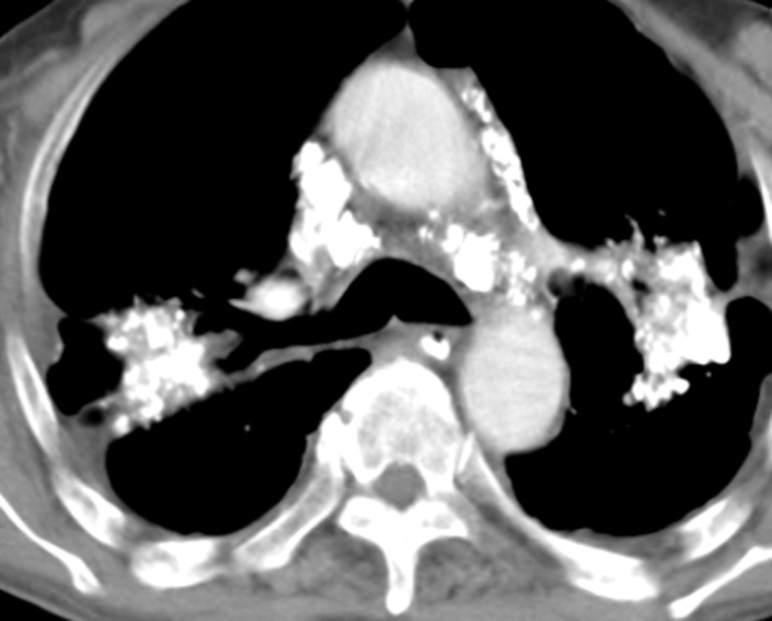
Progressive massive fibrosis in a 61-year-old man. Frontal chest radiograph (a) shows large bilateral upper lobe–predominant masses with irregular margins (arrows) and upper lobe volume loss, indicated by a juxtaphrenic peak (arrowhead). (b) Axial CT image shows bilateral large calcified conglomerate masses with adjacent fibrosis and calcified mediastinal lymph nodes.