. 2020 Jun 5;40(4):1003–1019. doi: 10.1148/rg.2020190180

Figure 19a.

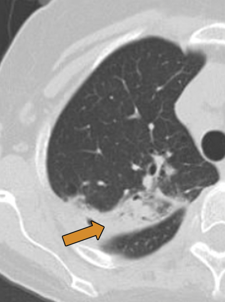
Biopsy-proven extranodal Rosai-Dorfman disease in a 65-year-old woman. (a–c) Axial CT images (lung window) show a patchy consolidation in the right upper lobe (arrow in a) with septal line thickening (arrow in b). Also note the tiny thin-walled cysts in the left lung (arrow in c). (d) Axial CT image (mediastinal windows) shows the axillary and mediastinal adenopathy (green arrows) and a soft-tissue chest wall mass (pink arrow). The most common finding in Rosai-Dorfman disease is lymphadenopathy. Most of these findings resolved with treatment (not shown).

Biopsy-proven extranodal Rosai-Dorfman disease in a 65-year-old woman. (a–c) Axial CT images (lung window) show a patchy consolidation in the right upper lobe (arrow in a) with septal line thickening (arrow in b). Also note the tiny thin-walled cysts in the left lung (arrow in c). (d) Axial CT image (mediastinal windows) shows the axillary and mediastinal adenopathy (green arrows) and a soft-tissue chest wall mass (pink arrow). The most common finding in Rosai-Dorfman disease is lymphadenopathy. Most of these findings resolved with treatment (not shown).