. 2020 Jun 5;40(4):1003–1019. doi: 10.1148/rg.2020190180

Figure 7b.

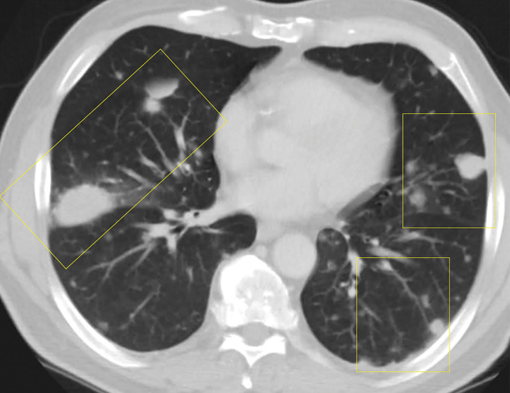
Lymphomatoid granulomatosis in a 54-year-old man who presented with shortness of breath and skin nodules. (a) Frontal chest radiograph shows multiple pulmonary nodules of varying sizes (yellow boxes). (b, c) Axial CT image (b) and coronal maximum intensity projection CT image (c) show these nodules (yellow boxes in b and c) as solid and distributed throughout the lung. (d) Captured rotating maximum intensity projection 18F fluorodeoxyglucose PET/CT image shows diffuse hypermetabolic activity of these pulmonary nodules. Multiple hypermetabolic cutaneous nodules are also seen (purple boxes). Skin biopsy results confirmed the diagnosis of lymphomatoid granulomatosis.

Lymphomatoid granulomatosis in a 54-year-old man who presented with shortness of breath and skin nodules. (a) Frontal chest radiograph shows multiple pulmonary nodules of varying sizes (yellow boxes). (b, c) Axial CT image (b) and coronal maximum intensity projection CT image (c) show these nodules (yellow boxes in b and c) as solid and distributed throughout the lung. (d) Captured rotating maximum intensity projection 18F fluorodeoxyglucose PET/CT image shows diffuse hypermetabolic activity of these pulmonary nodules. Multiple hypermetabolic cutaneous nodules are also seen (purple boxes). Skin biopsy results confirmed the diagnosis of lymphomatoid granulomatosis.