. 2020 Jun 5;40(4):1003–1019. doi: 10.1148/rg.2020190180

Figure 9c.

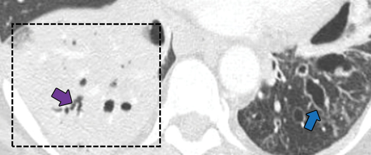
Bronchocentric granulomatosis in three patients. (a) Axial CT image in a 35-year-old man shows a spiculated nodule (arrow) in the left lower lobe. (b) Axial CT image in a 44-year-old woman shows peribronchovascular consolidation (white box) with tiny satellite nodules (arrow). (c) Axial CT image in a 51-year-old woman shows right lower lobe consolidation (dashed box) with peripheral bronchiectasis (purple arrow) and cystic bronchiectasis (blue arrow) with mucus plugging in the left lower lobe.

Bronchocentric granulomatosis in three patients. (a) Axial CT image in a 35-year-old man shows a spiculated nodule (arrow) in the left lower lobe. (b) Axial CT image in a 44-year-old woman shows peribronchovascular consolidation (white box) with tiny satellite nodules (arrow). (c) Axial CT image in a 51-year-old woman shows right lower lobe consolidation (dashed box) with peripheral bronchiectasis (purple arrow) and cystic bronchiectasis (blue arrow) with mucus plugging in the left lower lobe.