. 2020 May 15;40(4):1107–1124. doi: 10.1148/rg.2020190145

Figure 11b.

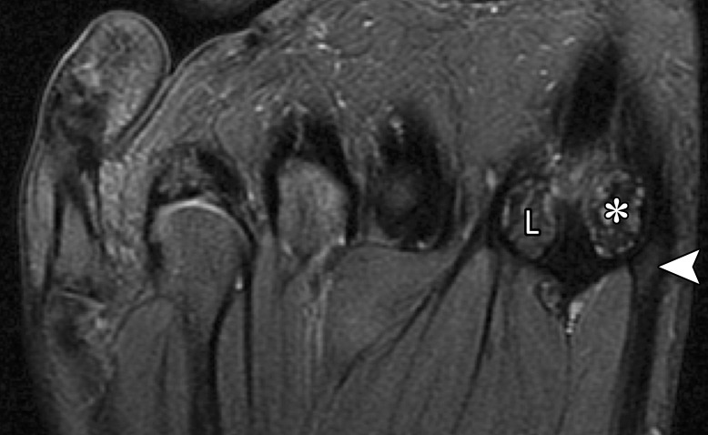
Medial MTSL tear in a 21-year-old man. Coronal (a) and axial (b) T2-weighted FS images show mild chondral loss (*) at the medial sesamoid and metatarsal head articulation. Partial tearing of the medial MTSL and capsular structures is seen with a fluid cleft at the sesamoid insertion (arrow). Thickening and altered signal intensity of the medial head of the FHB and abductor hallucis tendons (arrowhead) is compatible with tendinosis. These medial plantar structures interlink to prevent hallux valgus. L = lateral sesamoid, MT = first metatarsal.

Medial MTSL tear in a 21-year-old man. Coronal (a) and axial (b) T2-weighted FS images show mild chondral loss (*) at the medial sesamoid and metatarsal head articulation. Partial tearing of the medial MTSL and capsular structures is seen with a fluid cleft at the sesamoid insertion (arrow). Thickening and altered signal intensity of the medial head of the FHB and abductor hallucis tendons (arrowhead) is compatible with tendinosis. These medial plantar structures interlink to prevent hallux valgus. L = lateral sesamoid, MT = first metatarsal.