. 2020 May 15;40(4):1107–1124. doi: 10.1148/rg.2020190145

Figure 12b.

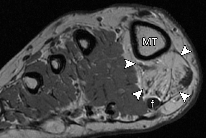
Lateral MTSL tear in a 54-year-old woman with right hallux valgus and first web space pain. Coronal T2-weighted FS (a) and PDW (b) images show chondral loss at the lateral sesamoid (*) and first metatarsal articulation (dashed oval). There is partial tearing of the lateral MTSL (☆) with adjacent first web space edema and adductor hallucis tendinosis (solid white arrow). Medial sagittal band tearing (dotted arrow) and mild lateral subluxation of the extensor tendons (black arrow) are due to underlying hallux valgus. There is atrophy of the abductor hallucis and the FHB muscles (arrowheads) due to long-standing hallux valgus deformity. f = flexor hallucis longus tendon, MT = first metatarsal.

Lateral MTSL tear in a 54-year-old woman with right hallux valgus and first web space pain. Coronal T2-weighted FS (a) and PDW (b) images show chondral loss at the lateral sesamoid (*) and first metatarsal articulation (dashed oval). There is partial tearing of the lateral MTSL (☆) with adjacent first web space edema and adductor hallucis tendinosis (solid white arrow). Medial sagittal band tearing (dotted arrow) and mild lateral subluxation of the extensor tendons (black arrow) are due to underlying hallux valgus. There is atrophy of the abductor hallucis and the FHB muscles (arrowheads) due to long-standing hallux valgus deformity. f = flexor hallucis longus tendon, MT = first metatarsal.