. 2020 May 15;40(4):1107–1124. doi: 10.1148/rg.2020190145

Figure 13b.

Lateral MTSL tear and adductor hallucis tendinosis and atrophy in a 69-year-old woman with hallux valgus and persistent foot pain. Coronal PDW (a, c) and T2-weighted FS (b) images show first MTPJ degenerative changes with chondral loss and subchondral edema most apparent at the lateral sesamoid (L) and metatarsal articulation. There is partial tearing of the lateral MTSL (dashed circle) and lateral collateral ligament (black arrow). More proximally in b, there is predislocation syndrome with edema surrounding the distal second (m) and third metatarsal shafts and MTPJs due to capsulitis. Tendinosis of the transverse (solid white arrow) and oblique (dotted arrow) heads of the adductor hallucis is apparent with fatty muscle atrophy (arrowheads) seen proximally in c. f = flexor hallucis longus tendon, M = medial sesamoid, MT = metatarsal.

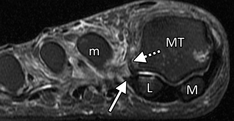
Lateral MTSL tear and adductor hallucis tendinosis and atrophy in a 69-year-old woman with hallux valgus and persistent foot pain. Coronal PDW (a, c) and T2-weighted FS (b) images show first MTPJ degenerative changes with chondral loss and subchondral edema most apparent at the lateral sesamoid (L) and metatarsal articulation. There is partial tearing of the lateral MTSL (dashed circle) and lateral collateral ligament (black arrow). More proximally in b, there is predislocation syndrome with edema surrounding the distal second (m) and third metatarsal shafts and MTPJs due to capsulitis. Tendinosis of the transverse (solid white arrow) and oblique (dotted arrow) heads of the adductor hallucis is apparent with fatty muscle atrophy (arrowheads) seen proximally in c. f = flexor hallucis longus tendon, M = medial sesamoid, MT = metatarsal.